Clinical Case Scenario
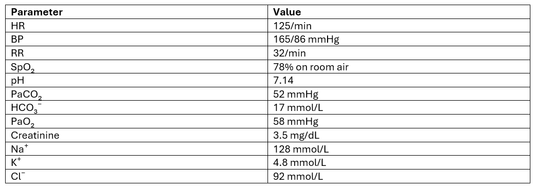
A 72-year-old man with diabetes mellitus, hypertension, coronary artery disease with PCI 10 years ago, HFrEF with an EF of 37%, CKD on medical management with a baseline creatinine of 1.8 mg/dL, and COPD requiring occasional home BiPAP presents with acute respiratory distress.
He has had progressively reduced mobility and bilateral lower-limb edema for the past 6 months. For the last 3 days, he has developed cough with expectoration associated with intermittent fever.
On examination, he is tachypneic with bilateral crackles, elevated JVP, and bilateral pitting pedal edema. Chest radiograph shows bilateral pulmonary edema.
Initial Assessment
Echocardiography shows LV systolic dysfunction with EF 37%, grade II diastolic dysfunction, mild RV dysfunction, moderate-to-severe tricuspid regurgitation, PASP approximately 52 mmHg, and a dilated IVC with no respiratory collapse.
POCUS demonstrates extensive bilateral B-lines involving all lung fields with small bilateral pleural effusions.
Initial VExUS
- IVC: Markedly dilated
- Hepatic vein: Severe abnormality
- Portal vein: Severe pulsatility
- Intrarenal vein: Severe abnormality
- Overall VExUS: Grade 3
The patient is diagnosed with AECOPD, likely precipitated by community-acquired pneumonia, with concurrent acute decompensated HFrEF. He is started on NIV, IV loop-diuretic infusion, and appropriate treatment for the suspected infective exacerbation. Over the next 48 hours, his respiratory distress and oxygen requirement improve considerably, with reduction in pulmonary edema.
Day 3
The patient is significantly more comfortable from a respiratory standpoint but continues to have elevated JVP and peripheral edema. Urine output has decreased and his response to further loop-diuretic doses is becoming poor.
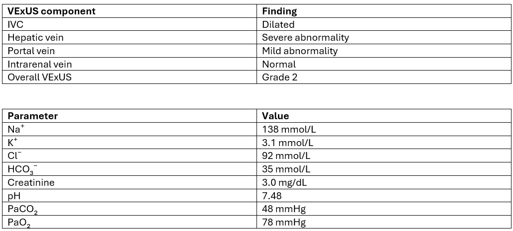
Question: What is the most appropriate next step in management?