Skip to content
Nephro Critical Care Society
  • Home
  • About
    • About Us
    • Our Journey
    • Vision, Mission & Objectives
    • Affiliations
  • Activities
    • Quiz OF The Week
    • Knowledge Sharing
    • Training Courses
    • Research
      • GLOBE RRT Survey
    • Literature
    • 5th Global Update In Nephro Critical Care POCUS IN NEPHRO CRITICAL CARE (PINC)
  • Blog
  • Media
    • Gallery
    • Videos
      • CRRT
      • AKI
      • Investigations & Biomarker
      • Fluid Therapy
      • Drug Dosing
      • Journal Scan
      • Other Extracorporeal Therapy
      • 5th Global Update In Nephro Critical Care POCUS IN NEPHRO CRITICAL CARE (PINC)
      • 5th Nephrocritical Care Review Course
  • Membership
    • Membership Benefits
    • All Members
    • Plus Members
  • My Account
    • Login
  • Contact Us
  • Home
  • About
    • About Us
    • Our Journey
    • Vision, Mission & Objectives
    • Affiliations
  • Activities
    • Quiz OF The Week
    • Knowledge Sharing
    • Training Courses
    • Research
      • GLOBE RRT Survey
    • Literature
    • 5th Global Update In Nephro Critical Care POCUS IN NEPHRO CRITICAL CARE (PINC)
  • Blog
  • Media
    • Gallery
    • Videos
      • CRRT
      • AKI
      • Investigations & Biomarker
      • Fluid Therapy
      • Drug Dosing
      • Journal Scan
      • Other Extracorporeal Therapy
      • 5th Global Update In Nephro Critical Care POCUS IN NEPHRO CRITICAL CARE (PINC)
      • 5th Nephrocritical Care Review Course
  • Membership
    • Membership Benefits
    • All Members
    • Plus Members
  • My Account
    • Login
  • Contact Us

QW64 July 2026

Question: Select each option to validate with explanations

Clinical Case Scenario

A 29-year-old previously healthy primigravida undergoes an emergency caesarean section for placental abruption at 37 weeks of gestation. The procedure is complicated by massive postpartum hemorrhage (estimated blood loss 5.2 L), profound hemorrhagic shock, and disseminated intravascular coagulation. She receives massive transfusion (12 units packed red blood cells, fresh frozen plasma, platelets, and cryoprecipitate), vasopressor support, and correction of coagulopathy.

On admission:

  • Hemoglobin: 5.5 g/dL
  • Platelets: 52 × 10⁹/L
  • INR: 2.8
  • Fibrinogen: 85 mg/dL
  • Lactate: 10.2 mmol/L
  • Serum creatinine: 0.8 mg/dL

Over the next 72 hours:

  • Hemodynamic status normalizes.
  • Vasopressors are discontinued.
  • Lactate- 1.3 mmol/L.
  • Coagulation profile normalizes.
  • Platelet count recovers to 105 × 10⁹/L.

Despite recovery from shock, she remains anuric (<20 mL/day), and serum creatinine increases to 7.4 mg/dL with hyperkalemia requiring intermittent hemodialysis.

Peripheral smear shows no schistocytes, LDH and bilirubin are within normal limits, and haptoglobin is normal. Renal ultrasonography demonstrates normal-sized kidneys without hydronephrosis. Urinalysis: Specific gravity 1.010, trace protein, 5–10 RBC/HPF, 1–2 WBC/HPF, no muddy brown granular casts or renal tubular epithelial cell casts, and no RBC casts.

Day 6

On the sixth postoperative day, the patient develops progressive abdominal distension, diffuse abdominal pain, nausea, and failure to pass flatus, raising concern for postoperative ileus, bowel ischemia, intra-abdominal collection, or bowel injury. Physical examination reveals a distended abdomen with diffuse tenderness but no peritoneal signs.

A contrast-enhanced CT scan of the abdomen and pelvis is performed to evaluate the cause of the acute abdomen.

CECT Abdomen: Dilated small- and large-bowel loops consistent with paralytic ileus, with no evidence of bowel perforation or intra-abdominal abscess. Kidneys “Reverse Rim sign”
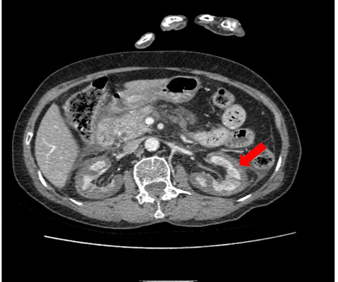
Image courtesy: https://radiopaedia.org

Question: Which of the following is the most likely diagnosis?
😭

Wrong Answer: ❌ A. Rapidly Progressive Glomerulonephritis (RPGN)
RPGN typically presents with dysmorphic RBCs, RBC casts, significant proteinuria, and often systemic autoimmune features, which are absent in this patient.

😭

Wrong Answer: ❌ B. Acute Tubular Necrosis (ATN)
ATN is common after hemorrhagic shock, but ATN usually shows recovery of urine output after restoration of perfusion and is characterized by muddy brown granular casts and renal tubular epithelial cell casts, none of which are present.

😉

Right Answer: ✅ C. Acute Cortical Necrosis (ACN)

Explanation

The combination of:
• Severe postpartum hemorrhage
• DIC
• Prolonged shock
• Persistent anuria for 6 days despite restoration of hemodynamics
• Progressive rise in creatinine
• CT findings
is highly suggestive of acute cortical necrosis.

Acute cortical necrosis
Acute cortical necrosis (ACN) is a rare but severe cause of intrinsic AKI characterized by ischemic necrosis of the renal cortex, while the medulla is relatively spared due to its separate blood supply. Unlike acute tubular necrosis (ATN), cortical necrosis is often irreversible.

Etiology
Obstetric causes (most common worldwide)
• Placental abruption
• Postpartum hemorrhage
• Septic abortion
• Preeclampsia/eclampsia
• HELLP syndrome
• Amniotic fluid embolism

Non-obstetric causes
• Septic shock
• Severe trauma or burns
• Disseminated intravascular coagulation (DIC)
• Hemolytic uremic syndrome (HUS)
• Snake envenomation
• Acute pancreatitis
• Massive hemorrhage
• Rarely, severe vascular injury or transplant rejection

Pathophysiology
• Severe systemic hypotension and endothelial injury
• Intrarenal vasospasm and microvascular thrombosis
• Cortical ischemia leading to coagulative necrosis
• Medulla is usually preserved

Clinical features
• Sudden severe AKI
• Persistent oliguria or anuria
• Gross hematuria may occur
• Flank pain (occasionally)
• Features of the underlying disorder (e.g., DIC, sepsis, obstetric catastrophe)

Confirmation
• Contrast CT kidney: cortical non-enhancement with preserved medullary enhancement (“reverse rim sign”)
• MRI: Demonstrates cortical infarction.
• Renal biopsy (gold standard if diagnosis remains uncertain): Patchy or diffuse cortical necrosis. Usually reserved when diagnosis remains uncertain.

Prognosis
• ATN → usually reversible
• ACN → often results in permanent loss of renal function, CKD, or dialysis dependence
table

Key Learning Point
• Suspect ACN in persistent anuric AKI after obstetric catastrophes, severe septic shock, DIC, or snakebite. Failure of renal recovery after several days to weeks despite correction of the initial insult.
• The characteristic contrast-enhanced CT finding is cortical non-enhancement with preserved medullary enhancement (Reverse Rim Sign).

CLINICAL PEARLS

💎 Persistent anuria after severe PPH and DIC
🎯 Think Acute Cortical Necrosis.
🎯 Reverse Rim Sign on contrast CT is a characteristic imaging clue.
🎯 Renal biopsy remains the gold standard when imaging is inconclusive.

😭

Wrong Answer: ❌ D. Acute Interstitial Nephritis (AIN)
AIN is usually drug-induced and presents with fever, rash, eosinophilia, sterile pyuria, or WBC casts, features not seen in this case.

This field is for validation purposes and should be left unchanged.
Nephro Critical Care Society

All rights reserved