Adj. Associate Professor – National University Heart Centre, Singapore
Adult Cardiac Intensivist | ECMO Educator | ELSO Scientific Co-Chair
A 63-year-old male is admitted to hospital with acute decompensated heart failure secondary to anterior wall MI. He has a history of hypertension and diabetes and is on treatment for the same. His vitals are:
• BP: 100/75 mm Hg; HR: 90 bpm; CVP ~15 mm Hg; peripheral edema (+)
• S. creatinine 1.7 mg/dL; Na 131
• POCUS: LVEF 35%, Ascites and bilateral pleural effusions (+)
• Treatment: IV nitrates and diuretics
Day 1: fluid balance is 2L. Patient remained stable after PCI to LAD.
Day 2: UO 0.4 ml/kg/hour for 5 hours.
Patient remained on 2L oxygen via nasal prongs. His S.creatinine increased to 2.7 mg/dL; peripheral edema persisted.
(Click / Tap on Questions to Reveal Content)
Cardiorenal Syndrome Type 1 (CRS-1) is a condition where acute worsening of heart function leads to acute kidney injury (AKI). It is seen in patients with acute decompensated heart failure (ADHF), where the heart’s inability to pump effectively results in decreased renal perfusion and subsequent kidney dysfunction.
The development of CRS-1 involves a complex interplay between the heart and kidneys:
——————————————————————————————————————-
Patients with CRS-1 typically present with:
——————————————————————————————————————-
Effective management of CRS-1 requires a multifaceted approach:
1. Hemodynamic Stabilization
2. Renal Support
Renal Replacement Therapy: In severe cases, dialysis may be required to manage fluid overload and electrolyte disturbances.
3. Monitoring and Supportive Care
The European Society for Cardiology 2021 guideline considers intravenous diuretics as the ‘cornerstone of acute heart failure management’ even though the level of evidence is low (‘C grade’). It is a clinical myth that that the action of frusemide is often ascribed to the diuresis that follows an intravenous bolus. The sight of a full urine bag is considered as a token of successful treatment that reduces the work of breathing.
However, the benefit from intravenous furosemide arises from its rapid vasodilating effect. Changes in cardiac loading conditions with a 27% fall in left ventricular filling pressure and a 52% increase in mean calf venous capacitance within a few minutes has been established
minutes following a furosemide bolus. The use of diuretics in acute LVF always remains an important dogma in cardiac critical care.
Many question the rationale for using furosemide as first-line therapy in patients with acute heart failure who are not intravascularly overloaded. Nevertheless, the mechanism of frusemide action in acute LVF can be summarised as :
——————————————————————————————————————-
Frusemide has a rapid venodilatory effect, even before significant diuresis occurs:
——————————————————————————————————————-
Frusemide is a loop diuretic that acts on the thick ascending limb of the loop of Henle in the kidney.
——————————————————————————————————————-
By reducing blood volume, frusemide decreases venous return (preload) to the heart.
——————————————————————————————————————-
——————————————————————————————————————-
In acute LVF with pulmonary edema, IV frusemide is preferred because of:
However, frusemide should be used carefully, as excessive diuresis or too rapid fluid shifts can:
Worsening renal function (WRF) during the treatment of cardiorenal syndrome occurs in approximately one third of heart failure admissions. This has its repercussions on length of stay, readmission rate, and short- and long-term survival. It is imperative that diuresis causes loss of intravascular fluid volume and this impacts on kidney function eventually. Many physicians would stop diuretics in this context. However, studies have shown that continuing aggressive diuresis in WRF leads to haemoconcentration and is associated with positive impact on survival. Patients who get discharged from hospital with elevated filling pressures have a subsequent higher risk of mortality. As such active decongestion would be required to disrupt the pathophysiological processes implicated in heart failure. Ironically similar correlations between PAC derived cardiac filling pressures and cardiorenal interactions are sparse highlighting the fact that pressure has little correlation with volume in the venous system. In such instances haemoconcentration and protein concentration during active diuresis seem better markers of the efficacy of diuresis as well as of outcomes.
Diuretic resistance is the inability to achieve euvolemia despite increasing doses of diuretic therapy. The diagnosis needs assessment of volume status and evaluation of possible contributing factors.
In general loop diuretics block a major site of sodium reabsorption, the Na-K-2Cl co- transporter (NKCC2) in the thick ascending loop of Henle and macula densa. As a result, sodium goes along the tubule taking water along with resulting in diuresis as well as natriuresis. When diuretic response is lost in heart failure two things may happen: (a) there might be insufficient delivery of loop diuretics to the proximal tubule, and (2) increased sodium hunger leading to sodium reabsorption at other nephron segments.
It can be challenging to diagnose diuretic resistance. A spot urine sodium collected two hours after administering a diuretic can help predict whether or not natriuresis will be adequate and can facilitate early recognition of an impaired diuretic response. It is however
difficult to differentiate whether this is due to insufficient loop diuretic delivery or distal sodium reabsorption

There are different ways of approaching diuretic response. As a preliminary step, it might be prudent to alter the route of loop diuretic administration (IV vs oral) or increasing the dose/ frequency.

It is important to understand some of the terminologies associated with diuretic resistance in this context. The “ceiling dose” of a diuretic refers to the single dose amount that achieves diuretic efficiency; doses above this ceiling do not meaningfully increase diuresis. The usual ceiling dose is about 80 mg of Frusemide, increased doses of oral diuretics can be required in the setting of impaired kidney drug delivery, with doses as high as 5 times being required. It is generally accepted that once the ceiling dose has been identified for an individual, this dose can be given up to every 6 hours to maximize the total daily urine output. The ceiling dose can change in an individual over time.
Apart from Furosemide, Torsemide and Bumetanide are becoming increasingly popular first
line choices due to improving costs and beneficial clinical profiles. The bioavailability is 80%-100% and are more effective than oral furosemide.
For a rough bedside conversion:
40 mg furosemide PO = 1 mg bumetanide PO/IV (most potent) = 20 mg torsemide PO/IV (longest duration of action)
One way to alleviate diuretic resistance is to reduce distal Na reabsorption. Thiazide and thiazide-like diuretics block the Na-Cl symporter in the distal convoluted tubule and the NDCBE-pendrin system in the collecting ducts. As such loop diuretics can be combined with thiazides to block the proximal as well as distal Na absorption sequentially and treat diuretic resistance – a concept referred to as “sequential nephron blockade”.
A combination regimen including four medications — a loop diuretic, thiazide, acetazolamide, and spironolactone — is called “multi-nephron segment diuretic therapy”. Multi-nephron segment diuretic therapy works at different segments of the nephron and appears to be effective in treating diuretic resistance without significant electrolyte abnormalities
Furosemide’s effectiveness and safety are closely tied to kidney function. Since it’s mostly eliminated through the urine and partly metabolized in the kidneys, advanced CKD can lead to higher drug levels and an increased risk of ototoxicity—sometimes irreversible. In contrast, bumetanide and torsemide are processed in the liver and may be safer in severe kidney dysfunction.
In hospitalized patients, IV loop diuretics are often used at 2.5 times the home oral dose to ensure adequate effect. A spot urine sodium test (goal >50 mmol/L) 1–2 hours after dosing can help assess response and guide further dosing using validated prediction tools. If the response is poor, a higher dose can be given immediately.
Continuous IV infusion of loop diuretics hasn’t clearly outperformed intermittent dosing in trials. Dosing decisions in ASCEND-HF trial were made by treating physicians- the findings reflect real-world practice: sicker patients tended to receive infusions. In situations requiring large diuretic doses, clinicians often preferred continuous infusions to avoid high serum peaks and potential ototoxicity—though evidence supporting this approach remains limited.
While an infusion may slightly increase urine output and be preferred in sicker patients to avoid serum peaks, strong evidence supporting its benefit is limited.
In cases of poor kidney perfusion, improving blood flow can enhance diuretic response. Inotropes (e.g., dobutamine), vasoconstrictors (e.g., midodrine), and renal vasodilators (e.g., low-dose dopamine, nesiritide) can help depending on the underlying issue
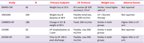
Unfortunately, there are situations in which all the diuretics in the world won’t effectively mobilize fluid. UF is the removal of isotonic fluid via convection, typically achieved using a dialysis machine. It is often considered in patients who have acute congestion refractory to multiple diuretics strategies, or those who have severe AKI or advanced CKD limiting diuretic efficacy. UF can be a short-term strategy used to support volume removal pending kidney recovery. However, there is limited data supporting its widespread use. In CARRESS-HF, UF was inferior to a stepped pharmacologic treatment strategy. UF has been associated with harm; those who were randomized to receive UF in CARRESS-HF and in the AVOID-HF
trial experienced significantly more adverse effects than those on pharmacologic treatment. The following table summarises the outcomes of UF trials in CRS 1.


Personalized medicine has the potential to significantly improve outcomes in CRS-1 by tailoring treatment based on individual patient characteristics, biomarkers, and pathophysiology.
WRF plus reduced NP = No WRF plus reduced NP ➔ Prognosis good at hospital discharge
No WRF plus no reduction NP = intermediate outcomes WRF plus no reduction/ increased NP = worse outcomes
![]()